Bone: Periosteal chondroma
2012-05-01 Nilo Sakai Junior, PhD , Ricardo Karam Kalil Affiliation1.The Sarah Network of the Rehabilitation Hospitals, Surgical, Molecular Pathology Laboratories, Brasilia\/DF, Brazil
Summary
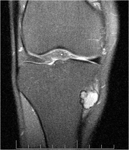
Figure 1: MRI S/DP SPIR - This coronal view shows a hyperintense lobulated lesion on the anteromedial surface of the tibial metaphysic (courtesy of Dr. Ricardo Karam Kalil).
Classification
Classification
Cartilaginous tumors of bone.
Clinics and Pathology
Phenotype stem cell origin
Mesenchyme.
Embryonic origin
Probably from the cambium layer of the periosteum.
Epidemiology
Periosteal chondromas comprise less than 1% of bone neoplasms, have preference for the first 3 decades of life, although it can be seen in any age and men predominate over female patients 3:2. It usually affects the metaphysis of long bones, by far predominating in the femur, tibia or humerus.
Clinics
Most frequently a swelling, sometimes associated to slight pain are the usual symptoms. Imaging show a well-delimited uniformly lucent lesion on the surface of bone situated over a saucerized, sclerotic depression of the cortex.
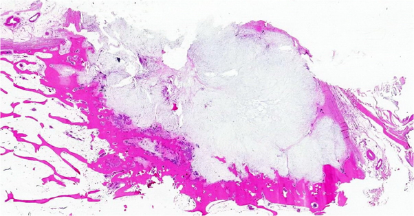
Figure 2: Tissue section, whole mount of the lesion, HE. This panoramic view of the same case as fig. 1 shows a bluish, cartilaginous lesion on the surface of bone, with sclerotic margins in the inner face, and partly covered by an external fibrous membrane (courtesy of Dr. Ricardo Karam Kalil).
Pathology
Grossly, it is a well-circumscribed, rubbery, lobulated nodule, measuring less than 3cm in its greatest dimension, with a membranaceous periosteal covering over the external surface, and in direct contact with a dense bone cortex on its inner face.
Microscopically, it is a hyaline cartilage lobulated tumor; chondrocytes may be enlarged, hyperchromatic and with double nuclei, eliciting a histological differential diagnosis with surface chondrosarcoma and parosteal osteosarcoma. Focal myxoid changes may be seen.
Microscopically, it is a hyaline cartilage lobulated tumor; chondrocytes may be enlarged, hyperchromatic and with double nuclei, eliciting a histological differential diagnosis with surface chondrosarcoma and parosteal osteosarcoma. Focal myxoid changes may be seen.
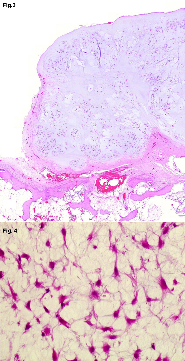
Figure 3: Tissue section, HE, 20X - Mature lobular cartilaginous tissue constitutes the usual finding in periosteal chondromas (courtesy of Dr. Ricardo Karam Kalil). Figure 4: Tissue section, HE, 40X - Occasional lesions may show myxoid cartilaginous areas. Same case as fig. 1 (courtesy of Dr. Ricardo Karam Kalil).
Treatment
Complete local surgical excision.
Evolution
Periosteal chondroma is a slow growing tumor, rarely surpassing 3cm in its greatest dimension. If the lesion exceeds 5cm a serious possibility of malignancy must be considered.
Prognosis
Excellent. Rare recurrences are cured by re-excision.
Cytogenetics
Note
Cytogenetics studies of periosteal chondromas are scarse. A total of 7 cases with abnormal karyotypes have been reported (Table 1). No consistent abnormality has been detected, although we observed one case of a periosteal chondroma with a t(2;11)(q37;q13) (Sakai et al., 2011). This alteration was previously described in one enchondroma (Dahlen et al., 2003). Recently, Amary and colleagues reported that 56% of central and periosteal cartilaginous tumors contain somatically acquired, heterozygous mutations in isocitrate dehydrogenase 1 (IDH1) or IDH2 (Thomas, 2011). They identified 5 periosteal chondromas with IDH1/IDH2 mutation type (71.43%). IDH1/IDH2 mutations result in elevated levels of HIF-1a and the associated transcriptional activity of its target genes, and that these effects are mediated through low levels of a-ketoglutarate (Zhao et al., 2009). These genes are important in adaptation of cells to low oxygen tension and are involved in glucose metabolism, angiogenesis, cell motility and invasion functions that are important for tumor growth/progression.
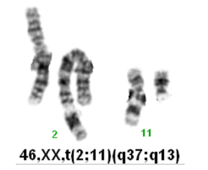
Figure 5: Karyotype image of periosteal chondroma with t(2;11)(q37;q13) (courtesy of Dr. Nilo Sakai Junior).
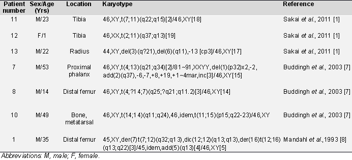
Table 1: Cytogenetic data for periosteal chondroma (Mitelman et al., 2012).
Article Bibliography
Citation
Nilo Sakai Junior ; Ricardo Karam Kalil
Bone: Periosteal chondroma
Atlas Genet Cytogenet Oncol Haematol. 2012-05-01
Online version: http://atlasgeneticsoncology.org/solid-tumor/5334/bone-periosteal-chondroma
